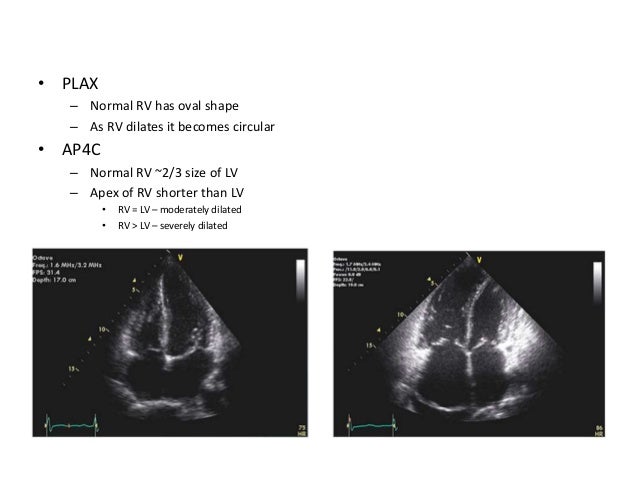
Rv Lv Ratio Echocardiography
Https Encrypted Tbn0 Gstatic Com Images Q Tbn 3aand9gctrtldus7mggg4dy8o1fwklwf3yeeeu 8qcng Usqp Cau
Basic Haemodynamic Assessment With Echo Iheartscan
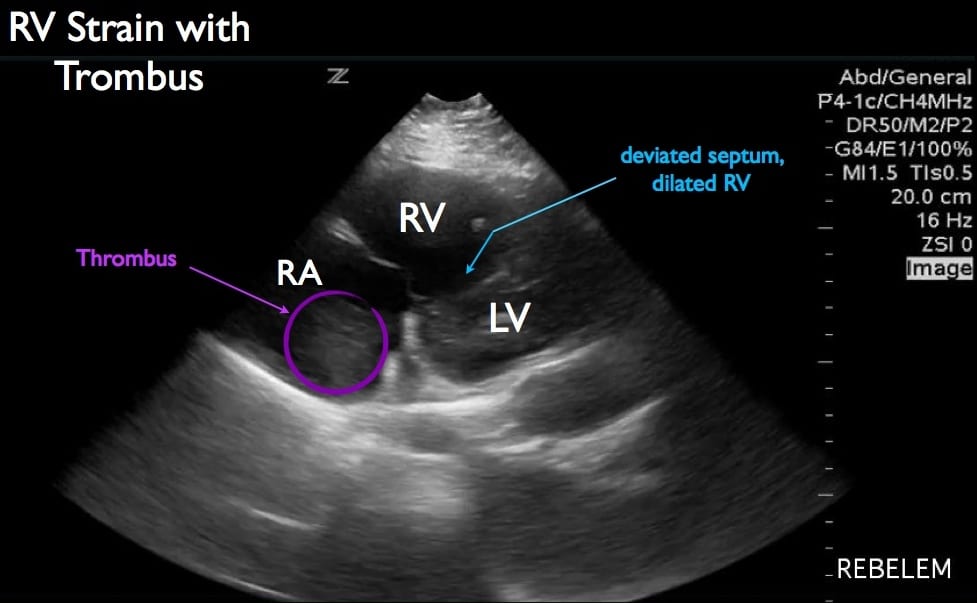
Diagnosis Of Right Ventricular Strain With Transthoracic Echocardiography Rebel Em Emergency Medicine Blog

Figure 1 From Increased Right To Left Ventricle Diameter Ratio Is A Strong Predictor Of Right Ventricular Failure After Left Ventricular Assist Device Semantic Scholar

Imaging Right Left Ventricular Interactions Jacc Cardiovascular Imaging

Acep American College Of Emergency Physicians
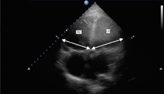
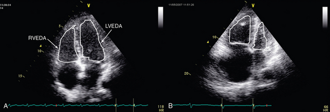
Right heart strain or more precisely right ventricular strain is a term given to denote the presence of right ventricular dysfunction usually in the absence of an underlying cardiomyopathy.
Rv lv ratio echocardiography.

Ctpa Demonstrating The Rv Lv Ratio Measurement Note Ctpa Computed Download Scientific Diagram
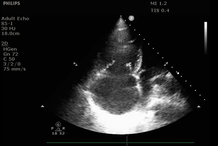
Differentiating Acute Versus Chronic Right Heart Failure With Bedside Echocardiography Emra
Submassive Pe Emory School Of Medicine
Evaluation Of Right Ventricular Function In The Intensive Care Unit By Echocardiography Consultant Level Examination Radiology Key
Source : pinterest.com
